Angiosarcoma is a rare type of cancer that represents only about 0.1% to 0.2% of all breast cancers. In the year 2020, the total occurrence of breast sarcoma will rise from 1.52 to 2.04 million cases per year. This cancer occurs in the breast and the arms' skin and starts to develop in the blood or lymphatic vessels in the breast. Angiosarcoma can grow and spread throughout the body very rapidly. They are rare and make up less than 1 in 100 breast cancers. It is generally seen in women but is also common in men. Its varied types include primary angiosarcoma and secondary angiosarcoma. The former starts in the breast tissue and consists of the skin of the breast. They are most likely to develop in younger women in their 30s or 40s. The secondary angiosarcomas of the breast occur because of undergoing radiotherapy to the breast for previous breast cancer. These cancers typically develop in older women. This condition gets a bit difficult to treat. The common symptoms of breast angiosarcoma include:
- a change in the shape of the nipple, mainly if it turns in, sinks into the breast, or has an irregular shape
- a lump or thickening in an area of the breast
- a rash or bruising on the breast or nipple
- a change in the size or shape of a breast, dimpling of the skin
- a swelling or lump in the armpit
With regard to the diagnostic procedures involved in this diseased condition, several techniques are used to diagnose these patients. Tests such as a mammogram, an ultrasound, especially if you are under the age of 35 years, a biopsy, or an MRI scan. Effective treatments are very much necessary to make a recovery faster. Breast angiosarcomas are very rare; thus, there is no set standard treatment. Many patients are recommended for mastectomy, which means breast removal. Surgical oncologists at many breast centers specialize in breast conservation surgery and can provide patients with advanced techniques in mastectomy and reconstruction of the breast. Then few patients with abnormal lymph nodes in the armpit (axilla) are recommended a sentinel lymph node biopsy. Chemotherapy or targeted drugs are always an option. Radiotherapy might not be an option if you previously had breast cancer radiotherapy. In recent times, for women with breast cancer or with a positive BRCA mutation who are willing to undergo prophylactic mastectomy, there is a rapidly growing demand for breast reconstruction with the use of breast implants. Patients undergoing breast implants tend to be concerned that the presence of a mass may go unobserved, and the diagnosis may be delayed.
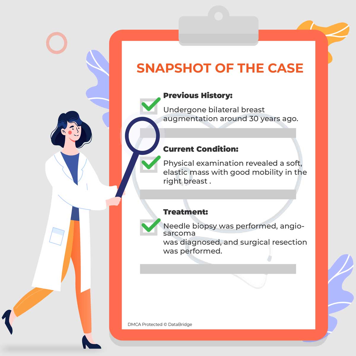
This case reports an older woman who had undergone bilateral breast augmentation around 30 years ago who came to the clinic complaining about a right breast mass. Surgical resection was performed following a core needle biopsy that showed pathological features of angiosarcoma. The patient had a good postoperative course and has remained recurrence-free for 4 years.
Case Description:
An 83-year-old female presented to the clinic with a right breast mass.
Diagnostic Tests Performed:

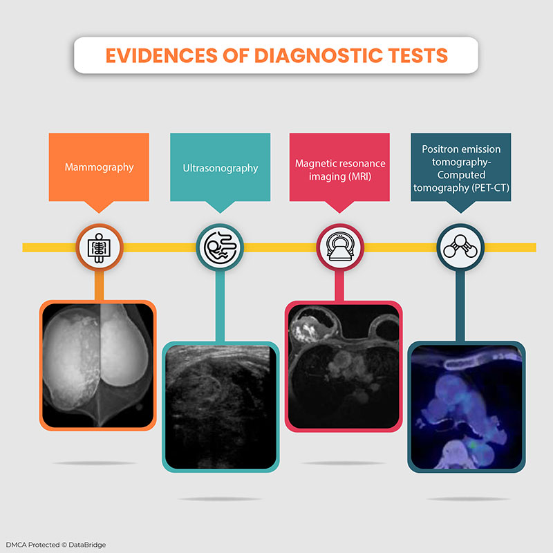
Mammography: A dense, oval, marginated mass over the bilateral breasts was observed. Another oval-shaped, marginated mass with coarse internal calcification was witnessed adjacent to this mass in the right breast.
Ultrasonography: Mass shadow with rough margins and heterogeneous internal structure on the dorsal side of the implant.
Magnetic Resonance Imaging (MRI): The posterior gap of the right breast implant revealed a heterogeneous high-to-low-signal mass lesion on T2-weighted images and an intermediate signal mass lesion on T1-weighted images.
A granular lesion was witnessed within that showed substantial enhancement from the early stage of contrast enhancement, and the dynamic curve revealed a rapid washout pattern.
Positron Emission Tomography-Computed Tomography (PET-CT): Mild hyperaccumulation was witnessed in the right breast with a maximum standardized uptake value of 1.80, which was consistent with the mass.
Core Needle Biopsy: Nuclear enlargement, stratification, microvessel anastomoses, and channel formation.
Besides the diagnostic factors, immunostaining was performed for keratin A/E 1/3, factor VIII, D2-40, and CD68 was negative and indicated that the tumor was mostly derived from endothelial cells.
Observations During Surgery:
Nipple-sparing mastectomy was performed. The tumor and implant were covered by a capsule. No tumor invasion was seen into the dermis, and the major pectoralis muscle was witnessed.
Pathological Reports:
The total size of the removed specimen was 8 × 6 × 13 cm. A size 6 × 4 × 7 cm mass was found dorsal to the implant. The tumor was dark red grossly, revealing lesions with organizing thrombus formation and large and small cyst-like cracks.
A magnified image revealed the presence of mammary tissue between the tumor and the capsule. Histopathological analysis revealed that more than 75% of the tumor consisted of low-grade, less atypical areas. Still, scattered substantial growth was observed, giving an intermediate appearance between low and high grades.
The tumor was diagnosed to be an intermediate-grade angiosarcoma rising from the mammary tissue between the implant and capsule.
Treatment Post Surgery:
The patient was discharged on the fourth-day post-operation. She was advised of no adjuvant chemotherapy and later was under regular follow-up. No recurrence was observed even after almost 4 years of surgery.
Key Highlights:
- Breast angiosarcoma is a malignant tumor with the mean age of onset for the primary type is 59 years, and that for the secondary type of 72.9 years
- Different imaging techniques are helpful in diagnosing the diseased condition
- In this case, it was noticed that the tumor was covered by the implant and was difficult to palpate; hence, in this regard, multiple imaging is necessary
- Surgical resection is considered to be the first-line treatment for patients with breast angiosarcoma
- Here, diagnostic tests such as MRI, ultrasonography, mammography, Positron emission tomography-Computed tomography (PET-CT), core needle biopsy were performed to diagnose the condition
- In implant-related breast angiosarcoma, the rate of recurrences is higher and the prognosis may be poor, but in this case, the patient witnessed no recurrences till date
Conclusion:
Breast angiosarcoma is not so common, and there is no treatment for it. However, surgical resection is chosen to be the primary treatment. The age of onset range from 31 to 87 years and many cases occurred at least 20 years after implant insertion. It has been studied and witnessed that implant-related breast angiosarcoma is not age-related, although it takes a long time to develop. Patients should be provided with appropriate diagnostic procedures to make the treatment process faster.